Thèse: équipe Lumière, vision et cerveau
Titre: Utilisation de CRISPR-Cas9 chez Psammomys obesus pour examiner la vulnérabilité des cônes aux mutations du gène Abca4 impliqué dans la maladie de Stargardt.
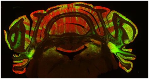
Résumé: La maladie de Stargardt 1 (STGD1) est une maladie autosomique récessive. Le gène dans lequel les mutations sont responsables a été identifié, Abca4, codant pour un membre de la super famille des cassettes de liaison à l’ATP, exprimé spécifiquement dans la rétine. L’objectif de ce projet est de comparer la vulnérabilité des photorécepteurs en bâtonnets et cônes à la perte d’Abca4 par une stratégie CRISPR-CAS9 utilisant un rongeur diurne. Le modèle animal, Psammomys obesus, est une espèce diurne riche en cônes dans laquelle les cônes sont représentés en grand nombre (~ 30-40% de photorécepteurs totaux), ressemblant plus étroitement à la macula humaine et permettant des détails structurels, fonctionnels et analyses biochimiques. Nous avons effectué des injections sous-rétiniennes de constructions Adeno-Associated Virus (AAV)-CRISPR-Cas9 chez Psammomys obesus, pour obtenir un Abca4 – KO. Une fois ayant confirmé la perte d’Abca4, pour analyser les conséquences fonctionnelles et morphologiques nous avons réalisé des analyses non invasives (électrorétinographie, tomographie par cohérence optique, imagerie du fond d’œil). Les yeux traités présentent une dégénérescence rétinienne étendue et rapide 2 mois après l’injection, comme le montre l’OCT, et l’enregistrement ERG a montré une baisse significative des réponses lumineuses photopiques (cônes). En conclusion, les rongeurs riches en cônes offrent un scénario unique pour explorer les changements moléculaires et cellulaires survenant dans les maculopathies humaines telles que STGD1, et devrait fournir un moyen précieux d’évaluer les stratégies thérapeutiques potentielles.
Title: Use of CRISPR-Cas9 in Psammomys obesus to examine the vulnerability of cones to mutations in the Abca4 gene involved in Stargardt disease.
Summary: Loss of central vision constitutes the major underlying reason for visual handicap in western industrialized nations. Stargardt’s Disease 1 (STGD1) is an autosomal recessive disease of early onset and severe visual handicap, representing the most frequent inherited macular degeneration. The gene in which mutations are responsible for STGD1 has been identified, Abca4, coding for a member of the ATP binding cassette super family, expressed specifically in retina. The aim of this project is to compare the vulnerability of rod and cone photoreceptors to Abca4 loss by a CRISPR-Cas9 strategy using a diurnal rodent. The animal model, Fat Sand Rat Psammomys obesus, is a diurnal cone-rich species in which cone photoreceptors are represented in large numbers (~30-40% total photoreceptors), more closely resembling the human macula, and allowing detailed structural, functional and biochemical analyses. We performed subretinal injections of Adeno-Associated Virus (AAV)-CRISPR-Cas9 constructs in Psammomys obesus, to obtain an Abca4 – KO. After confirming the loss of Abca4; to analyse the functional and morphological consequences to Abca4 loss we performed non-invasive imaging (Electroretinography, Optical coherence tomography, Fundus imaging). CRISPR-Cas9-Abca4 -/- treated eyes exhibit extensive and rapid retinal degeneration by 2 months post-injection, as seen by OCT, and ERG recording showed significant declines in photopic (cone) light responses.In conclusion, cone-rich rodents offer a unique scenario to explore molecular and cellular changes occurring in human maculopathies like STGD1, and should provide a valuable means to appraise potential therapeutic strategies.
Position actuelle : Post-doctorat
Institut: Institut des Neurosciences Cellulaires et Intégratives, UPR-3212, Strasbourg, France
Team leader (chef d’équipe): David Hicks

Upright microscope equipped for observation in bright light and epifluorescence.